Penn Quantitative Imaging Resource for Pancreatic Cancer
Here we provide the software used to analyze the pre-clincal and patient data
Pre-clininical and clinical MR imaging datasets are provided along with manually defined lesion masks
Genetically engineered mouse (GEM) models are used to study the development of pancreatic ductal adenocarcinoma.
Protocols on animal modes, imaging, and processing.
Description: This is a command line tool that allows for the conversion of Varian files (typically named with the .fdf extension) to be converted to more commonly used image formats such nifti
Description:This dataset includes baseline and post-treatment diffusion-weighted MRI (DW-MRI) and dynamic contrast-enhanced MRI (DCE-MRI) from a pancreatic cancer patient enrolled in clinical trial entitled “A Randomized Pilot Study of Perioperative Nivolumab and Paricalcitol to Target the Microenvironment in Resectable Epithelial Subtype Pancreatic Cancer” (NCT03519308).
Reference:
Description: Test-Retest Diffusion imaging in 10 mice. Includes original data as well as derived images such as: ADC, M0, M0 standard deviation, BL, and BL standard deviation.
Reference: Cao J, Song HK, Yang H, Castillo V, Chen J, Clendenin C, Rosen M, Zhou R, Pickup S. Respiratory Motion Mitigation and Repeatability of Two Diffusion-Weighted MRI Methods Applied to a Murine Model of Spontaneous Pancreatic Cancer. Tomography. 2021; 7(1):66-79. https://doi.org/10.3390/tomography7010007
Resource Type: Image Data Site: Github.com Link: MouseDiffusionImaging Research Area: Diffusion Imaging
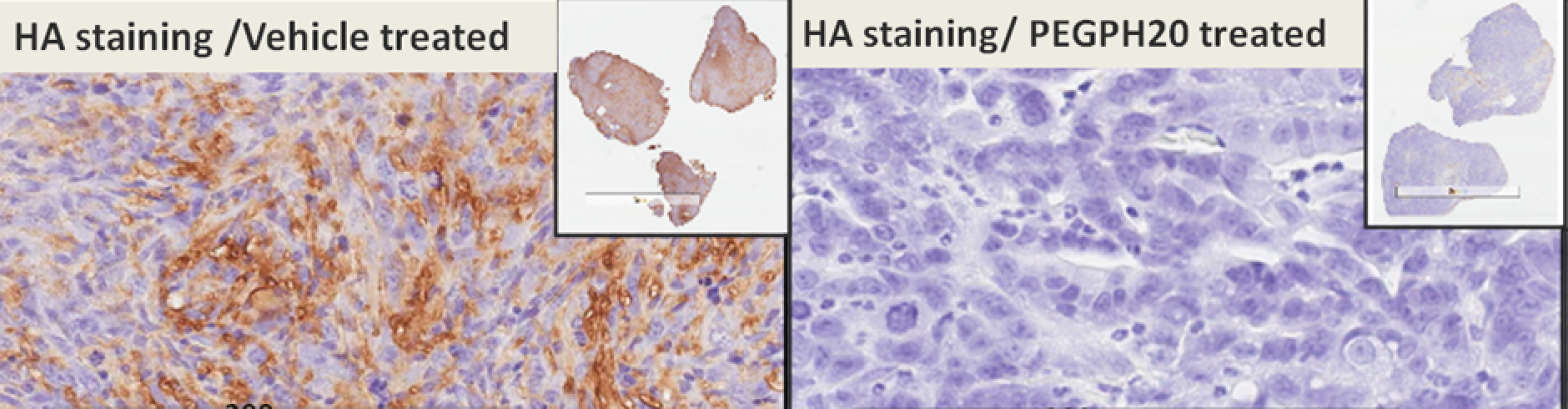
Description: PDA development is accompanied by the formation of a unique tumor microenvironment, i.e., a dense and complex stroma that includes fibroblasts, blood vessels, and immune cells. It is well known that pancreatic cancer stroma is best modeled in autochthonous tumors, which arise spontaneously from the native locations as the result of genetic mutations. Therefore, to study stromal interventions, the genetically engineered mouse (GEM) models are strongly preferred.
Mice harboring a pancreas specific Kras and p53 mutant with Cre alleles (KPC) were initially developed at Penn (Reference Paper) and is bred at the Mouse Hospital of Penn Pancreatic Cancer Research Center (For more information, download the Overview PDF).
Besides the GEM model, we have established orthotopic allograft model and xenograft (human PDA cell line) model. Both models are described in a Clinical Cancer Research paper, which compares the DCE-MRI results from KPC, orthotopic allograft and xenograft models.
Resource Type: GEM Model Site: Github.com Link: Overview (pdf) Research Area: Pancreatic cancer
Description: Acquisition of Diffusion MRI of Mouse Abdomen
Description: Processing of Diffusion MRI of Mouse Abdomen
Description: Acquisition of dynamic contract enhanced MRI of mouse abdomen
Description: Processing of dynamic contract enhanced MRI of mouse abdomen